To cite this section of the Cancer Epigenetics Drug Database

ReferenceS. Winkler, A. Neudolt, T. Habenschuss, and I.M. Bennani-Baiti. DNA methyltransferase inhibitors. Cancer Epigenetics Drug Database (CEDD) – Experimental dataset, Cancer Epigenetics Society (https://ces.b2sg.org/cedd/experimental_dnmti/), 2019.

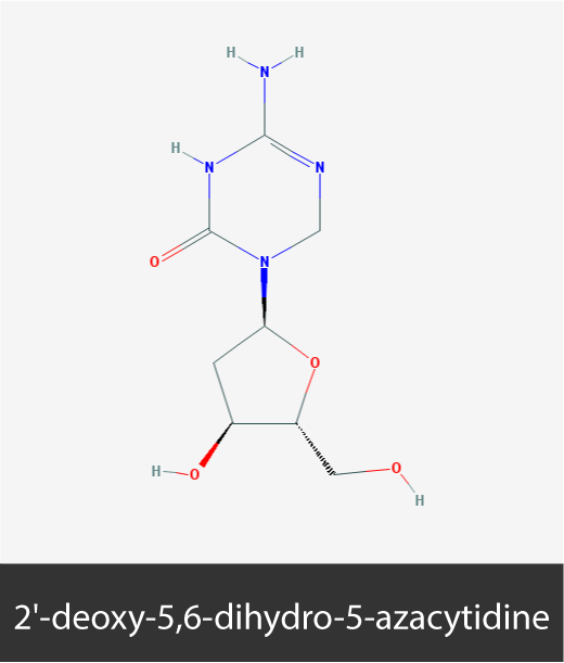
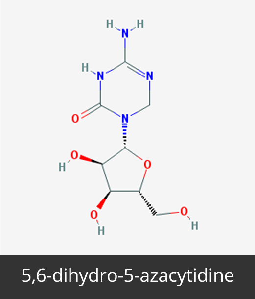
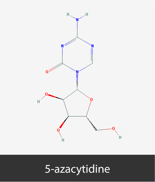
Chemical structures of cancer epigenetic drugs

Brief description of cancer epigenetic drugs

0
Drugs
0
Cell lines
0
Experiments

2′-deoxy-5,6-dihydro-5-azacytidine is a DNA methyltransferase (DNMT) suicide substrate that can be integrated into DNA, leading to DNA hypomethylation and expression reactivation of silenced genes.

2′-deoxy-5,6-dihydro-5-azacytidine is water and DMSO soluble (up to 100 mM), and 2′-Deoxy-5,6-dihydro-5-azacytidine solutions can be stored at -20C for up to a month with little loss of activity.

2′-deoxy-5,6-dihydro-5-azacytidine chemical structure is 4-Amino-3,6-dihydro-1-β-D-ribofuranosyl-1,3,5-triazin-2(1H)-one hydrochloride. It is also known as 114522-16-6, 5,6-dihydro-5-aza-2′-deoxycytidine, KP 1212, SureCN3117083.

5,6-dihydro-5-azacytidine, a derivative of 5-azacytidine and a deoxycytidine analog, is a DNA methyltransferase inhibitor (DNMTi). 5,6-dihydro-5-azacytidine is thought to exert anticancer activity through the demethylation and reactivation of tumor suppressor genes silenced by DNA hypermethylation in tumors.

Molecular Weight: 246.223 g/mol.

5,6-dihydro-5-azacytidine is soluble in Water (<1 mg/ ml at 20°C), DMSO (≥ 100 mg / ml @ 20°C), and 95% ethanol( ≥ 100 mg/ ml @ 20C). Samples stored at 60C for 30 days display a < 1% degradation (TLC, UV).

5,6-dihydro-5-azacytidine is also known as dihydro-5-azacytidine 4-Amino-5,6-dihydro-1-beta-D-ribofuranosyl-s-triazin-2(1H)-one and as DHAC.

5-aza-2′-deoxycytidine is the deoxy derivative of 5-azacytidine. Its synthesis was first reported alongside that of 5-azacytidine in the 1960s, at which time it’s antineoplastic activity on cancer cell lines was also demonstrated. Today, 5-aza-2′-deoxycytidine is used to treat myelodysplastic syndrome (MDS). Similarly to 5-azacytidine, 5-aza-2′-deoxycytidine also incorporates into DNA, inhibits CpG DNA methylation, and arrests cells in the S phase of the cell cycle.

5-aza-2′-deoxycytidine chemical structure is 4-amino-1-(2-deoxy-beta-D-erythro-pentofuranosyl)-1,3,5-triazin-2(1H)-one and is also referred to as 5-aza-dCyd, DAC, deoxyazacytidine, or dezocitidine, and is traded under the brand names of Dacogen® and Decitabine®.

5-azacytidine is a cytidine analogue anti-cancer drug first synthesized in 1964 and currently used to treat myelodysplastic syndrome (MDS). 5-aza-cytidine incorporates into the DNA and reversibly inhibits de novo DNA methylation. 5-aza-cytidine can also incorporate into RNA and hampers tRNA cytosine-5-methyltransferase activity. Tumor suppressor genes silenced in tumors by DNA hypermethylation can be reactivated by 5-aza-cytidine.

5-azacytidine chemical structure is 4-amino-1-beta-D-ribofuranosyl-1,3,5-triazin-2(1H)-one and is also referred to in the literature as 5-AC, 5-azacytidine, AZA-CR, azacytidine, or ladakamycin, and is is traded under the names of Mylosar® and Vidaza®.

5-fluoro-2′-deoxycytidine is a fluorinated pyrimidine analogue prodrug that is metabolized into 5-fluoro-2-deoxyuridine monophosphate (FdUMP) and 5-fluorouridine triphosphate (FUTP). FdUMP blocks DNA synthesis (and therefore the cell cycle) by inhibiting thymidylate synthase and the production of thymidine monophosphate. FUTP also blocks cell proliferation by competing with uridine triphosphate (UTP) for incorporation into RNA, which in turn inhibits RNA and protein synthesis. 5-fluoro-2′-deoxycytidine inhibits DNA methyltransferases (DNMTs) by forming a covalent link with cysteine residues in their catalytic site.

5-fluoro-2′-deoxycytidine chemical structure is 4-amino-5-fluoro-1-[(2R,4S,5R)-4-hydroxy-5-(hydroxymethyl)tetrahydro-furan-2-yl]pyrimidin-2-one and is also referred to as 2′-deoxy-5-fluoro-Cytidine, FCDR, or FdCyd.

Epigallocatechin Gallate is an antioxidant flavonoid found in many plants, especially in dried white and green tea leaves, and to a lesser extent in black tea. It inhibits telomerase and DNA methyltransferases and may serve as a cancer chemopreventive agent.

Molecular weight: 458.372 g/mol.

EGCG is soluble at 10 mM in water or DMSO, and at 100 mM in ethanol.

EGCG is also known as Epigallocatechin-3-gallate or Tea catechin and its chemical structure is (2R,3R)-5,7-Dihydroxy-2-(3,4,5-trihydroxyphenyl)chroman-3-yl 3,4,5-trihydroxybenzoate.

Genistein is phytoestrogen and isoflavone that can be extracted from soybeans and fava beans. Genistein modulates the activity of a wide range of molecules, for example activating peroxisome proliferator-activated receptors (PPARs) and inhibiting several tyrosine protein kinases, DNA topoisomerase-II, and DNA methyltransferases. Genistein also displays antioxidant and antiangiogenic activities.

Genistein chemical structure is 5,7-dihydroxy-3-(4-hydroxyphenyl)-4H-1-benzopyran-4-one or 4′,5,7-trihydroxyisoflavone, and is also referred to as genisteol, genisterin, prunetol, or sophoricol.

Hydralazine is a vasodilator and hypotensive drug prescribed in cardiovascular conditions under the brand name of Apresoline® and Nepresol®. Hyralazine is also a DNA methyltransferase inhibitor, albeit weaker than other nucleoside analogues such as 5-azacytidine and 5-aza-2′-deoxycytidine, which display more favorable free binding energies for DNMT1. Consistent with its demethylating activity, hydralazine reactivates tumor suppressor gene expression and inhibits tumor cell proliferation.

Hydralazine is also referred to in the literature as 1-hydrazinophthalazine, Apressin, hydralazine, hydralazine hydrochloride, hydralazine mono-hydrochloride, Hydrallazin, hydrazinophthalazine, hydrochloride hydralazine, hypophthalin, and mono-hydrochloride hydralazine. Hydralazine is soluble in water at a concentration of 44.2 g/l at 25 C (30.1 g/l at 15 C).

Parthenolide, a sesquiterpene lactone of the germacranolide class, is the active principle of feverfew (Tanacetum parthenium). It depletes HDAC1 without affecting other class I/II HDACs, and induces DNA damage and apoptosis.

Molecular weight: 248.322 g/mol

Parthenolide is soluble at 100 mg/ml in DMSO, ethanol (up to 30 mg/ml), but is quasi-insoluble in water. Dimethylaminoparthenolide (DMAPT) is a water-soluble parthenolide analogue bioactive against several cancer cell lines.

Parthenolide chemical structure is (1aR,4E,7aS,10aS,10bS)-1a,5-Dimethyl-8-methylene-2,3,6,7,7a,8,10a,10b-octahydrooxireno[9,10]cyclodeca[1,2-b]furan-9(1aH)-one, and is also known as (-)-Parthenolide or Tanacetum parthenium.

Procainamide is a local and regional anesthetic, and a supraventricular and ventricular antiarrhythmic aminobenzamide drug prescribed under the trade names of Pronestyl®, Procan®, and Procanbid®. Procainamide efficiently inhibits DNMT1 activity on hemimethylated DNA substrate (Ki = 7.2 ± 0.6 μM), whereas it is ineffective in inhibiting DNMT1 activity on unmethylated DNA, or DNMT3a and DNMT3b on both hemimethylated and unmethylated DNA (1,400 < Ki <10,000 μM). Procainamide was reported to reactivate epigenetically inactivated tumor suppressor genes such CDKN2A, retinoic acid receptor beta (RARB), and Wnt inhibitory factor-1 (WIF-1).

Procainamide chemical structure is para-amino-N-2-(diethylamino)ethyl-benzamide and is water soluble at ~ 3 mg/ml.

Procaine is a benzoic acid derivative also known as Novocaine, Vitamin H3, Duracaine, or Spinocaine which possesses antiarrhythmic properties, constricts blood vessels, and exerts a local anesthetic activity (particularly used in oral/dental surgery). It is prescribed under the trade name of Novocain®. Procaine was reported to reactivate epigenetically inactivated tumor suppressor genes such retinoic acid receptor beta (RARB) and Wnt inhibitory factor-1 (WIF-1).

Procaine chemical structure is 4-Aminobenzoic-acid-2-diethylaminoethyl-ester and is water soluble at ~ 9.4 mg/ml (at 30C).

RG108 is a non-nucleosidic DNMT1 inhibitor that blocks DNMT1 catalytic site (IC50 = 115 nM) and induces significant genomic DNA demethylation, but not that of centromeric satellite DNA sequences. It also causes reactivation of tumor suppressor genes.

Molecular weight: 334.331 g/mol

RG108 is soluble at 100 mM in DMSO or ethanol.

RG108 chemical structure is (2S)-2-(1,3-dioxoisoindol-2-yl)-3-(1H-indol-3-yl)propanoic acid and is also known as N-Phthalyl-L-tryptophan.

S-equol is a dietary non-steroidal estrogen metabolite of the isoflavonoid daidzein. S-equol displays chemoprotective properties, suppresses dihydrotestosterone (DHT) production, and inhibits androgen-dependent prostate cancer cell growth.

Molecular weight: 242.274 g/mol

S-equol is soluble up to 100 mM in DMSO or ethanol.

S-equol chemical structure is (3S)-3-(4-hydroxyphenyl)-3,4-dihydro-2H-chromen-7-ol.

SGI-110 is a second generation DNA methyltransferase inhibitor (DNMTi) dinucleotide antimetabolite that consists of a deoxyguanosine (5’-DACpG-3’) linked to decitabine via a natural phosphodiester bond. Unlike other DNMTi, SGI-110 is not a substrate of cellular cytidine deaminase, which may extend the cellular half-life of Decitabine. Phosphorylation of SGI-110 leads to metabolic activation and incorporation into the DNA, which in turn leads to epigenomic hypomethylation and cell cycle arrest in the S phase.

SGI-110 chemical structure is [(2S,3R,5R)-5-(2-amino-6-oxo-3H-purin-9-yl)-3-hydroxyoxolan-2-yl]methyl [5-(4-amino-2-oxo-1,3,5-triazin-1-yl)-2-(hydroxymethyl)oxolan-3-yl] hydrogen phosphate, and is also referred to as MG98 or Guadecitabine. SGI-110 is highly insoluble and it is therefore recommended to dissolve it at 10 mM in DMSO at 37C under sonication.

Zebularine, a 2-H pyrimidinone-containing cytidine analog, forms covalent complexes with DNA methyltransferases and causes global DNA demethylation. Zebularine is a hydrophilic, stable in aqueous solutions, has good oral bioavailability, and it displays preferential targeting of cancer cells.

Molecular weight: 228,20 g/mol

Soluble up to 100 mM in water and up to 100 mM in DMSO

Zebularine chemical structure is 1-(beta-D-ribofuranosyl)-3,4-dihydropyrimidin-2(1H)-one and is also known as 3,4-Dihydrozebularine.

Functional assays used to assess the activity of cancer epigenetic drugs

Annexin V Assay is a cell viability assay that relies on the premise that Annexin V, a cell-impermeable protein, binds with high affinity membrane phosphatidylserine phospholipids when exposed at the cell surface. Though phosphatidylserine phospholipids localize to the inner, cytoplasmic side of the plasma membrane in healthy cells, they translocate from the inner to the outer side of the plasma membrane early in apoptosis, and become accessible to Annexin V. Following staining with Annexin V and propidium iodide (see PIA), the assay allows to quantify: i. (Annexin V / PI) viable cells; ii. Annexin V+ / PI cells undergoing early apoptosis; iii. (Annexin V / PI)+ dead cells and those in late apoptosis. Annexin V is usually labeled with a fluorochrome such as FITC as to analyze staining by flow cytometry.

Automated Cell Counting regroups a number of automated procedures including for example electrical impedance-based technologies (e.g Bio-Rad’s Coulter), or microscopy imaging-based methods (e.g. Beckman’s Vi-CELL™ Cell Counter) to assess cell viability and proliferation. These techniques are mostly automated versions of the trypan blue exclusion assay (see TBE).

BrdU Cell Incorporation Assay is a cell proliferation assay used to quantify 5-bromo-2′-deoxyuridine or BrdU incorporation into DNA during DNA replication that occurs in the course of cellular proliferation. Cells in culture are incubated in the presence of large amounts of BrdU, which is incorporated into DNA instead of thymidine. Following cell fixation and DNA denaturation, a labelled monoclonal anti-BrdU antibody is used to quantify the amount of incorporated BrdU, which in turn reflects the extent of de novo replicated DNA / cell proliferation.

Caspase 3/7 Apoptosis Assay: the Caspase 3 and 7 cysteine protease family members are integral components of mammalian apoptosis. Several variants of this assay are available, including for example those that utilize caspase 3 and 7 substrates that emit light or fluorescence once cleaved by the caspases. The substrate being in excess, the amount of light/fluorescence is proportional to the amount of caspases, and by extension, to the number of apoptotic cells.

Calcein AM allows to assess cell metabolism, and consequently, cell viability. Calcein AM is a non-fluorescent, cell permeable hydrophobic compound that is hydrolyzed by intracellular esterases to produce Calcein, a hydrophilic, cytoplasm-contained fluorescent compound in metabolically active (live) cells. Spectrophotometry of Calcein AM-labeled cells with an excitation at 490 nm and emission at 520 nm permits the quantification of metabolically active cells.

Cell Cycle Analysis is carried out by flow cytometry analysis of cellular DNA content after staining of nuclei with a DNA binding dye (usually propidium iodide). Cells distributes into the G0/G1 phase (cells with about 1n DNA content), S phase (~1.5n), and G2/M phase (~2n). This assay allows to assess (and quantify) if a drug induces a cell cycle block at a given cell cycle phase/checkpoint.

Colony Formation Assay or Colony Forming Units is an assay that is usually carried out by visualizing colonies with the crystal violet dye (see CVA) and allows to assess a cell’s capacity to overcome contact inhibition by growing into a colony, usually in soft agar. This assay is considered to be an in vitro measure of cellular tumorigenicity. The assay is based on counting the number of colonies, their size, or both.

Crystal Violet Assay is a cell viability and proliferation assay based on crystal violet, a cell permeable tri-(dimethylaniline)-methane dye that binds to both DNA and proteins, thus allowing for the visualization of cells (e.g. see CFA), or their quantification by use of spectrophotometry at 570 nm. Since crystal violet stains equally well living and dead cells, this assay is appropriate only for adherent cells that detach after cell death. After the medium is discarded and the dead cells washed away, only the adherent cells remain and are stained.

CyQUANT is a cell proliferation assay that relies on a cell permeable green fluorescent nucleic acid dye (CyQUANT GR dye) and a fluorescence suppression dye that quenches background cellular fluorescence. This is a one-step assay whereby reagents are added directly to cells in culture, incubated 30-60min, and fluorescence is measured with an excitation at 485 ± 10 nm and emission at 530 ± 12.5 nm. The assay essentially measures the amount of nucleic acids in living cells in culture, and is linear over a range of 50 – 250,000 cells in a 200 µl volume.

4′,6-diamidino-2-phenylindole or DAPI is a fluorescent dye that stains the nucleus by binding to AT-rich DNA. Apoptosis can be assessed in fluorescent microscopy by counting the number of DAPI-stained cell nuclei that show condensed and fragmented chromatin, two early signs of apoptosis.

Erythrosine B Exclusion assay is an alternative to trypan blue (see TBE) in quantifying cell viability and proliferation. Similarly to trypan blue, live cells are impermeable to Erythrosine B, whereas dead cells readily incorporate it.

CellTiter-Glo Luminescent Cell Viability Assay assesses the levels of cellular ATP as a gauge of cellular metabolism. The assay is based on luciferase, which requires ATP (and Mg2+) to convert luciferin into oxiluciferin, and which in the process emits luminescence. The intensity of luminescence signal is proportional to the amount of available cellular ATP, and is thus indicative of metabolic activity.

Guava Viability Assay allows to assess both total cell count and cell viability. The assay is based on quantifying two dyes, one that binds DNA in all cells, and a second dye that binds DNA in dead cells only.

Hemocytometer Cell Counting is based on counting the number of cells in visible light microscopy in multiple fields of a hemocytometer, and then calculating the average number of cells per field.

Lactate Dehydrogenase assay measures the amount of LDH, a non-secreted cytosolic enzyme, in cell culture media as an indicator of cytotoxicity. In cytotoxic conditions, the plasma membrane is damaged and LDH is released into the cell culture medium. The amount of LDH in the medium can be indirectly assessed using MTT type of assays that quantify the amount of NADH in the cell culture medium produced during the conversion of lactate into pyruvate by LDH.

Mitochondrial Membrane Potential assay: intact cells present with a mitochondrial membrane potential due to the polarity differential between the overall electronegatively charged mitochondrial matrix and electropositively charged cytosol. In this assay, cells are incubated in the presence of cell permeable potential-sensitive fluorescent dyes such as JC-1, which accumulate into mitochondria and shift fluorescence emission from green to red (from about 529 to 590 nm). In cytotoxic conditions or apoptosis, mitochondrial membranes are damaged and mitochondrial membrane polarity is compromised, leading to a shift in the fluorescence emission ratio, which can be quantified and reflects cell viability.

A variation of the MTT assay (see below) that uses the MTS reagent (3-(4,5-dimethylthiazol-2-yl)-5-(3-carboxymethoxyphenyl)-2-(4-sulfophenyl)-2H-tetrazolium) that also relies on formazan quantification to assess cellular metabolism, which may under certain conditions reflect cell number. Contrary to the multistep MTT assay, MTS involves a 1-step procedure.

Mosmann’s Thiazolyl blue Tetrazolium salt assay is a colorimetric assay to quantify cellular metabolic activity. MTT (3-(4,5-dimethylthiazol-2-yl)-2,5-diphenyltetrazolium bromide) is a yellow tetrazole that is metabolized into a non-hydrosoluble purple formazan by NADH/NADPH-dependent mitochondrial dehydrogenases. After solubilization in DMSO or SDS/HCl, formazan is spectrophotometrically quantified at 500-600 nm to assess the extent of tetrazole reduction, and by inference the activity of cellular dehydrogenases. Cellular metabolic activity may under certain conditions reflect cell number.

Neutral Red Method uses a vital dye, neutral red (a.k.a. Basic Red 5, Toluylene Red) that is actively uptaken by viable cells, whereby the dye accumulates in lysosomes, whereas non-viable cells remain impermeable to the dye. After washing out the excess of dye, the incorporated dye is released from the cells and is ionized following incubation in an acidified ethanol solution, which yields its characteristic red color. The amount of neutral red incorporated by the cells can then be quantified spectrophotometrically and is proportional to the number of viable cells.

Propidium Iodide (PI) Assay is a cell viability assay based on the fact that healthy cells are impermeable to PI. Under certain cytotoxic conditions and during apoptosis, cell membranes are compromised and show increased permeability to PI, which accumulates in the cell nuclei, wherein it intercalates into DNA. PI is fluorescent and can therefore be quantified by fluorescence microscopy or spectrophotometry. Since PI also binds RNA, cleaner results may be obtained by pretreating cells with RNAses.

Resazurin Conversion Assay is based on the reduction of the non-fluorescent resazurin reagent into fluorescent resorufin by cellular dehydrogenases, and thus serves to measure the level of cellular metabolism (or as some interpret it, to assess the number of metabolically active -and thus viable- cells).

Syto 60 Apoptosis Assay: Syto probes are cell permeable fluorescent compounds that bind to DNA and RNA. As cells undergo apoptosis, there is a caspase-dependent loss of fluorescence. The amount of fluorescence emitted by Syto is, therefore, proportional to the number of viable cells and inversely proportional to the number of apoptotic cells.

Spheroid Formation Assay is an assay that allows to assess a cancer cell’s capacity to grow in 3D, and thus to overcome contact inhibition. Cells are embedded in soft agar in the presence or absence of the drug, and spheroids are allowed to grow some 10-14 days depending on cell proliferation rates. The number of spheroids are counted at the end of the experiment for each test sample.

is a cell viability and proliferation assay that utilizes Sulforhodamine B, an anionic aminoxanthene dye that engages into electrostatic interactions with basic amino acids under moderately acidic conditions. The binding of Sulforhodamine B to cellular proteins is linearly dependent on the amount of proteins (and therefore cells), as measured spectrophotometrically at 560-580nm (after washing out the excess of non-bound dye and solubilization in SDS). Sulforhodamine B, however, permeates both living and dead cells, thus limiting the scope of the assay to adherent cells that detach after cell death.

Trypan Blue Exclusion Assay is based on counting in visible light microscopy the number of cells dyed in blue (trypan blue-permeable cells are cells whose membrane is no longer intact, which is indicative of cell death) and trypan blue-impermeable cells (viable cells). Trypan blue is also referred to as diamine blue or Niagara blue.

3H-Thymidine Incorporation assay allows to measure the amount of thymidine incorporated into de novo replicated DNA during cell proliferation. 3H-thymidine is added directly to the cell culture and permitted to incubate some 4-12 hours depending on cell division rates. Labelled DNA is next harvested onto glass fiber filter paper, which can be processed in a scintillation beta-counter. The amount of beta counts/amount of 3H-thymidine incorporated is proportional to the amount of replicated DNA and cell proliferation.

Terminal deoxynucleotidyl transferase (TdT) dUTP Nick End Labeling (TUNEL) allows the detection of DNA fragmentation that accompanies apoptosis. The assay takes advantage of the capacity of TdT to add labeled dUTPs to nicks in the fragmented DNA of cells undergoing apoptosis. The incorporation of dUTPs labeled with either a fluorochrome (e.g. fluorescein-dUTP) or a hapten (e.g. biotin-dUTP, BrdUTP) can be readily quantified and is proportional to the extent of apoptosis.

Transwell Assay, also referred to as the Boyden chamber transwell assay, can be used to quantify cellular motility, migration, chemotaxis, or invasion. The assay is based on the capacity of some cells to traverse a (usually 8 µm) porous membrane (motility / migration) that may in addition be coated with a basement membrane extract, Collagen I or IV, or Laminin I (invasion). The compartment to which the cells would migrate may contain a chemoattractant (e.g. SDF1α/CXCL12), thus allowing to assess chemotaxis. At the end of the experiments, cells that crossed the membrane are stained and counted.

The Wound Healing Assay, a.k.a Scratch Assay, is a technique to assess cell migration propensity in vitro. The assay relies on tracking cells movements on a slide onto which a gap (wound, scratch) is applied.  While this method is widely accepted as a cell migration assay, we at the Cancer Epigenetics Society have concerns about its validity as its results may be compromised by the proliferation, or lack thereof, of the cells under investigation.

Water-soluble Tetrazolium salts assays utilize water-soluble dyes that yield formazans with different absorption spectra as an alternative to the water-insoluble and single-spectrum reagents of the MTT assay. Assay based on WST-1 (2-(4-Iodophenyl)-3-(4-nitrophenyl)-5-(2,4-disulfophenyl)-2H-tetrazolium) and perhaps more so on WST-8 (2-(2-methoxy-4-nitrophenyl)-3-(4-nitrophenyl)-5-(2,4-disulfophenyl)-2H-tetrazolium) offer several advantages as they are reduced outside the cells, include 5-methyl-phenazinium methyl sulfate that serves as an electron transfer mediator, and yield a water-soluble formazan. Unlike MTT, WST assays are accompanied with lower cell toxicity, do not require a solubilization step, and emit stronger signals. WST assays allow to quantify cellular metabolism extracellularly. Cellular metabolic activity may under certain conditions reflect cell number.

A variant of the MTT, MTS and WST assays that relies on 2,3-bis-(2-methoxy-4-nitro-5-sulfophenyl)-2H-tetrazolium-5-carboxanilide as the tetrazole, and akin to WST, is reduced to a water-soluble formazan, thus eliminating the last solubilization step. XTT displays higher sensitivity and dynamic range than MTT, and as the latter, allows to quantify cellular metabolism. Cellular metabolic activity may under certain conditions reflect cell number.

A word about the Cancer Epigenetics Drug Database – DNMTi section

Welcome to the Cancer Epigenetic Drug Database – Experimental dataset for DNA methyltransferase inhibitors (DNMTi). The epigenetics drug database gives an overview of cancer cell lines sensitivity to DNMTi in various functional assays (see assays abbreviations below). The page starts off with the chemical structures of the epigenetic drugs, a succinct description of the epigenetic drugs and functional assays used to test them in cancer cell lines, followed by Tables summarizing the data. Some of the epigenetic drugs also have clinical activities which are summarized in this page. Please note that the epigenetic drug solubility values provided here are only to be used as a guide, and that solubility depends on several variables such as the purity of the solute and solvent used, as well as the temperature used. Similarly, the IC50 values provided herein can be highly dependent on the type of functional assay used and length of incubation of cells with the epigenetic drugs.

The Cancer Epigenetics Drug Database is a work in progress and we rely on our members and readers to bring to our attention data that ought to be included here. We, therefore, welcome any information that you may provide that would help us make this database as useful a tool for your research as possible. You may email us your suggestions at info@ces.b2sg.org, Subject line: CEDD – DNMTi, Experimental dataset. The Cancer Epigenetics Drug Database is periodically updated, so please check it again for new data that may be relevant to your research.

Notes:

1Inc: Incubation of the cells in the presence of the drug in days.
2Assay: Please see details on functional assays above.
3Gene vs protein expression: Symbols in plain text denote proteins (e.g. EZH2). Symbols in italics denote the mRNA (e.g. EZH2). Symbols in bold (e.g. EZH2) denote both the mRNA and the protein. ↑ indicates an increase, whereas ↓ indicates a decrease in mRNA or protein expression or in the levels of an epigenetic mark.

DNA methyltransferase inhibitor (DNMTi) activities in cancer cells

Cell lineIC50 (µM)Inc1Assay2Additional information3ID #
CCRF-CEM> 1003 XTT↓ 50% global 5meCpG, ↓ 70-80% 5meCpG at THBS-1 & CDKN2B
CCRF-CEM> 1003A5A↓ 50% global 5meCpG, ↓ 70-80% 5meCpG at THBS-1 & CDKN2B
CCRF-CEM> 1005A5A↓ 50% global 5meCpG, ↓ 70-80% 5meCpG at THBS-1 & CDKN2B
HL-60> 1003XTT

© CEDD, Cancer Epigenetics Society; all rights reserved, 2019. Data may be included in academic publications contingent on the citation of the reference on top of this page.

Cell line
IC50 (µM)
Inc1Assay2Additional information3ID #
CCRF-CEM103 XTT↓ 40-50% 5meCpG at THBS-1 & CDKN2B
CCRF-CEM> 1003A5A
CCRF-CEM> 1005 A5A
HL-60> 1003 XTT
L1210101.5ACC

© CEDD, Cancer Epigenetics Society; all rights reserved, 2019. Data may be included in academic publications contingent on the citation of the reference on top of this page.
Cell Line
IC50 (µM)Inc1Assay2Additional information3ID #
6-10B5 – 103MTT
6-10B~ 103CCA
A4573~ 0.13TBE 
A5492.52MTT
A549> 325GVA
A673< 0.13
TBE
CCRF-CEM13XTT↓ 60% global 5meCpG, ↓ 70-80% 5meCpG at THBS-1 & CDKN2B
CCRF-CEM< 13A5A
CCRF-CEM< 15A5A
CNE-15 – 103
MTT 
CNE-1~ 103
CCA
HCT116< 0.113CFA 
HL-600.73XTT
HLE> 14MTS
HT-29> 1003
MTT 
KG1~ 0.033TBE 
HuH6> 14MTS
HuH7> 14MTS
MCF7305
MTT 
MCF753HCC
MOLM-130.0097GLO
MV4-110.0297GLO
NCI-H12991<IC50<25GVA
NCI-H197512
MTT
NCI-H19750.55
GVA 
SKM-10.0167GLOSynergizes with HMTi EPZ-5676
SW48> 1003MTT 
TC-71> 0.13TBE

© CEDD, Cancer Epigenetics Society; all rights reserved, 2019. Data may be included in academic publications contingent on the citation of the reference on top of this page.
Cell line
IC50 (µM)Inc1Assay2Additional information3ID#
BON-1< 0.54ACC
CCRF-CEM13XTT
HCT116> 53MTT
B10042
HCT116< 0.4
13
CFA
B10042
HCT116~ 1015TBE
B10012
HeLa> 105MTT
B10054
HL-606.23XXT
HLE5 – 104RCA
B10063
HLF~ 54RCA
B10064
HuH7< 54RCA
MOLM-130.587GLO
MV4-111.57GLO
NALM-7< 104TBE
B10039
RPMI8226~ 0.53TBE
B10066
RPMI8266< 0.56TBE
B10066
SiHa> 105MTT
B10055
SKM-10.65-2.057GLOSynergizes with HMTi EPZ-5676

© CEDD, Cancer Epigenetics Society; all rights reserved, 2019. Data may be included in academic publications contingent on the citation of the reference on top of this page.

Cell line
IC50 (µM)
Inc1Assay2ID #
A549> 1.5
5
GVA 
HCT116< 0.053MTT
HepG2~ 0.053MTT
KYSE150~ 0.053MTT
NCI-H129915GVA
NCI-H1975~ 0.055GVA
U2 OS~ 103MTT

© CEDD, Cancer Epigenetics Society; all rights reserved, 2019. Data may be included in academic publications contingent on the citation of the reference on top of this page.
Cell line
IC50 (µM)
Inc1Assay2ID #
A54960<IC50<805
GVA 
HCT116< 1013CFA
NCI-H1299405GVA
NCI-H1975~ 405GVA

© CEDD, Cancer Epigenetics Society; all rights reserved, 2019. Data may be included in academic publications contingent on the citation of the reference on top of this page.

Cell line
IC50 (µM)
Inc1Assay2ID #
A549~ 482MTT 
DU-145~ 254MTT
MCF7~ 204MTT
MDA-MB-231~ 154MTT
MRC-5> 1002MTT
PC-3> 254MTT

© CEDD, Cancer Epigenetics Society & B² Scientific Group, Ltd. All rights reserved, 2016. Data may be included in academic publications contingent on the citation of the reference on top of this page.

Cell line
IC50 µM
Inc1Assay2Additional information3ID #
22Rv1> 40
14MTTDNMT1 & DNMT3a/3b
 
CaSki~ 401MTT
CaSki~ 1602MTT
CaSki160 – 3203MTT
CaSki403CCACells arrest in S phase
CRL2480> 1002MTT
CRL2480~ 1003MTT
DU-145< 2014MTTDNMT1 & DNMT3a/3b
HeLa~ 401MTT
HeLa~ 402MTT
HeLa1603MTT
HeLa~ 403CCACells arrest in S phase
HepG2> 1002MTT
HepG250 – 1003MTT
LNCaP~ 2014MTT
PC3> 4014MTT
SiHa~ 401MTT
SiHa~ 1602MTT
SiHa160 -3203MTT

© CEDD, Cancer Epigenetics Society; all rights reserved, 2019. Data may be included in academic publications contingent on the citation of the reference on top of this page.

Cell line
IC50 (µM)
Inc1Assay2ID #
1205 Lu7.9~ 1 PIA
1205 Lu3-6~ 1MMP
A37515.9~ 1PIA
A3756-12~ 1MMP
A3756-12~ 0.5WHA
A5494.34MTT
ACHN0.1 – 15MTS
ACHN< 31MTS
ARH77~ 1.83MTS
BT-20~ 210 – 12SFA
BT-20~ 101MTT
BT-2010~ 1PIA
BT-20< 101MMP
BxPC-362XTT
Cho-CK~ 173TBE
Choi-CK~ 153TBE
DU-145~ 20.25CFA
DU-1450.1 – 15MTS
DU-145< 31MTS
DU-145~ 2.23GLO
DU-145< 52TBE
DU-145CD44hi~3.52GLO
DU-145CD44lo~3.53GLO
H929~ 1.83MTS
HCT116~ 251MTT
Hep3B~ 63TBE
HepG2~ 7.23TBE
HN22~ 151TBE
HN22~ 151MTS
HN22< 51DAPI
HT-29~ 251MTT
HT-29~ 201MMP
HT-2974MTT
HT-29~ 221MTT
IM9 ~ 1.73MTS
JCK~ 173TBE 
KMH2 ~ 7 3HCC
L428~ 93HCC
LAPC4~ 33GLO
LAPC4CD44hi~ 43GLO
LAPC4CD44lo~ 4GLO
LS147T ~ 251MTT
MC-3 ~ 201TBE
MC-3 ~ 151MTS
MC-3  < 51DAPI
MCF7241MTS
MCF79.52MTT
MCF79.52SRB
MCF7> 61TTI
MCF7~ 141MTT
MCF7~ 102MTT
MCF7~ 151LDH
MCF7~ 102LDH
MCF79.5 2MTT
MDA-MB-23182MTT
MDA-MB-231~ 210 – 12SFA
MDA-MB-23110~ 1PIA
MDA-MB-231< 101MMP
MDA-MB-23182MTT
MDA-MB-2317.42SRB
MDA-MB-231< 61TTI
MDA-MB-236~ 510 – 12SFA
MDA-MB-236~ 101MTT
MDA-MB-23610~ 1PIA
MDA-MB-236< 101MMP
MIA PaCa-222XTT
MKN-28~ 4.34MTT
MKN-45~3.74MTT
MKN-45~ 12TUNEL
MKN-743.34MTT
MM1.S~ 2.23MTS
MM1.S~ 33MTS
MM1.R~ 23MTS
NCI-H9291 – 22BCI
NIH-OVCAR-3~ 101MTT
NIH-OVCAR-3~ 101NRM
NIH-OVCAR-3~ 101GSH
OUR-100.1 – 15MTS
OUR-10> 31MTS
OVCAR-3> 7.51MTT
OVCAR-3> 7.51NRM
Panc-182XTT
PC3~ 20.25CFA
PC3~ 43GLO
PC352TBE
PC3CD44hi~ 1.53GLO
PC3CD44lo~ 2.43GLO
PLC~ 103TBE
RC-K8~ 103HCC
RPMI 8226~ 6.53MTS
RPMI 82262 – 52BCI
RPMI 8226/Dox6~ 33MTS
SCK~ 103TBE
SiHa~ 131MTT
SiHa~ 82MTT
SiHa~ 141LDH
SiHa~ 92LDH
SK-OV-3> 7.51MTT
SUDHL-4~ 33HCC
SUDHL-4BclXL~ 7.53HCC
SUDHL-4puro~ 43HCC
SW48010 – 251MTT
SW620~ 151MTT
TE6716.54MTT
TEX< 2.511CFU
U266~ 4.83MTS
VCaP~ 43GLO
VCapCD44hi~ 63GLO
VCapCD44lo~ 63GLO
WM79318.3~ 1PIA
WM7936 – 12~ 1MMP
WM793~ 6~ 0.5WHA

© CEDD, Cancer Epigenetics Society; all rights reserved, 2019. Data may be included in academic publications contingent on the citation of the reference on top of this page.

Cell line
IC50 (µM)
Inc1Assay2ID #
A54920003MTS
HCT11620003MTS
MCF71000-20003HCC
MCF7> 305MTT
NCI-H4602000
3MTS

© CEDD, Cancer Epigenetics Society; all rights reserved, 2019. Data may be included in academic publications contingent on the citation of the reference on top of this page.

Cell lineIC50 (µM)
Inc1Assay2ID #
A5491000-20003MTT
BT-20> 10004TUNEL
HCT1161000-20003MTT
HCT116> 40013CFA
Huh6> 20004MTT
Huh71500<IC50<20004MTT
HLE1500<IC50<20004MTT
MCF7> 10004TUNEL
MCF710003HCC
NCI-H4601000-20003MTT

© CEDD, Cancer Epigenetics Society; all rights reserved, 2019. Data may be included in academic publications contingent on the citation of the reference on top of this page.

Cell line
IC50 (µM)
Inc1Assay2ID #
22Rv1~ 5010MTT 
A549> 5125GVA
DU-145> 20014 MTT
HCT116> 2013CFA
HCT116> 1015TBE
LNCaP~ 5010MTT
NALM-6> 1015TBE
NCI-H1299~ 2565GVA
NCI-H19755125GVA
PC-3> 20014MTT

© CEDD, Cancer Epigenetics Society; all rights reserved, 2019. Data may be included in academic publications contingent on the citation of the reference on top of this page.

Cell line
IC50 (µM)
Inc1Assay2ID #
MDA-MB-231> 3501
MTT
MDA-MB-231> 3502MTT
MDA-MB-2312523
MTT
T47D2281
MTT
T47D~ 1502MTT
T47D~ 1503MTT

© CEDD, Cancer Epigenetics Society; all rights reserved, 2019. Data may be included in academic publications contingent on the citation of the reference on top of this page.

Cell line
IC50 (µM)
Inc1Assay2ID #
A2780> 0.11TBE
A2780> 0.18SFA
Hep3B0.023MTT
Hep3B0.001-0.013CFA
HepG20.433MTT
HepG20.1-13CFA
SKOV3< 0.11TBE
SNU-38763.43MTT
SNU-3870.1-13CFA
SNU-3980.253MTT
SNU-3980.13CFA
SNU-4490.293MTT
SNU-4490.001-0.013CFA
SNU-475543MTT
SNU-4750.1-13CFA

© CEDD, Cancer Epigenetics Society; all rights reserved, 2019. Data may be included in academic publications contingent on the citation of the reference on top of this page.

Cell line
IC50 (µM)
Inc1Assay2Additional information3ID #
A549> 2505
GVA 
A549> 2003MTT
CCRF-CEM> 1003XTT↓ 40% 5meCpG at THBS-1 & CDKN2B
HCT116> 1013CFA
HCT11625<IC50<503MTT
HeLa40<IC50<603WST
HeLa50<IC50<1003MTT
HepG2400<IC50<6001WST
HepG2> 10003MTT
HL-60> 1003XTT
HT29> 2003MTT
HT29> 1003MTT
HuCCT1~ 2003WST
HuCCT1~ 10001WST
KKU-100> 10003WST
KKU-M156> 10003WST
KKU-M213> 10003WST
MCF7> 2003MTT
NCI-H1299> 2505GVA
NCI-H1975> 2505GVA
RKO25<IC50<503MTT
SW48> 1003MTT
SW480> 2003MTT
SW620> 2003MTT
TFK-1200<IC50<4003WST
TFK-1> 10001WST

© CEDD, Cancer Epigenetics Society; all rights reserved, 2019. Data may be included in academic publications the contingent on citation of the reference on top of this page.