To cite this section of the Cancer Epigenetics Drug Database

Reference: A. Neudolt, I. Hübscher and I.M. Bennani-Baiti. Lysine demethylase inhibitors. Cancer Epigenetics Drug Database (CEDD) – Experimental dataset, Cancer Epigenetics Society (https://ces.b2sg.org/cedd/experimental_kdmi/), 2016.

Chemical structures of cancer epigenetic drugs

Brief description of cancer epigenetic drugs

0
Drugs
0
Cell lines
0
Experiments

4SC-202 is a selective inhibitor of LSD1, HDAC1, HDAC2, and HDAC3. 4SC-202 leads to multiple nucleation centers, a collapsed mitotic spindle, and a G2/M cell cycle arrest of A549 NSCLC cells.

4SC-202 is poorly soluble in water or ethanol ( < 1 mg/ml) and is soluble in DMSO up to 89 mg/ml (198.87 mM). For in vivo studies, 4SC-202 is soluble at 10 mg/ml in DMSO:PEG 300:Tween 80:H2O (2%/30%/5%/63%).

4SC-202 chemical structure is (E)-N-(2-aminophenyl)-3-(1-(4-(1-methyl-1H-pyrazol-4-yl)phenylsulfonyl)-1H-pyrrol-3-yl)acrylamide. 4SC-202 is also known as KB-72935.

Also known as LSD1 inhibitor IV
Also known as LSD1 inhibitor II
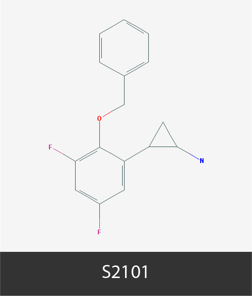
Also known as 2-PCPA

Functional assays used to assess the activity of cancer epigenetic drugs

Annexin V Assay is a cell viability assay that relies on the premise that Annexin V, a cell-impermeable protein, binds with high affinity membrane phosphatidylserine phospholipids when exposed at the cell surface. Though phosphatidylserine phospholipids localize to the inner, cytoplasmic side of the plasma membrane in healthy cells, they translocate from the inner to the outer side of the plasma membrane early in apoptosis, and become accessible to Annexin V. Following staining with Annexin V and propidium iodide (see PIA), the assay allows to quantify: i. (Annexin V / PI) viable cells; ii. Annexin V+ / PI cells undergoing early apoptosis; iii. (Annexin V / PI)+ dead cells and those in late apoptosis. Annexin V is usually labeled with a fluorochrome such as FITC as to analyze staining by flow cytometry.

Automated Cell Counting regroups a number of automated procedures including for example electrical impedance-based technologies (e.g Bio-Rad’s Coulter), or microscopy imaging-based methods (e.g. Beckman’s Vi-CELLâ„¢ Cell Counter) to assess cell viability and proliferation. These techniques are mostly automated versions of the trypan blue exclusion assay (see TBE).

BrdU Cell Incorporation Assay is a cell proliferation assay used to quantify 5-bromo-2′-deoxyuridine or BrdU incorporation into DNA during DNA replication that occurs in the course of cellular proliferation. Cells in culture are incubated in the presence of large amounts of BrdU, which is incorporated into DNA instead of thymidine. Following cell fixation and DNA denaturation, a labelled monoclonal anti-BrdU antibody is used to quantify the amount of incorporated BrdU, which in turn reflects the extent of de novo replicated DNA / cell proliferation.

Caspase 3/7 Apoptosis Assay: the Caspase 3 and 7 cysteine protease family members are integral components of mammalian apoptosis. Several variants of this assay are available, including for example those that utilize caspase 3 and 7 substrates that emit light or fluorescence once cleaved by the caspases. The substrate being in excess, the amount of light/fluorescence is proportional to the amount of caspases, and by extension, to the number of apoptotic cells.

Calcein AM allows to assess cell metabolism, and consequently, cell viability. Calcein AM is a non-fluorescent, cell permeable hydrophobic compound that is hydrolyzed by intracellular esterases to produce Calcein, a hydrophilic, cytoplasm-contained fluorescent compound in metabolically active (live) cells. Spectrophotometry of Calcein AM-labeled cells with an excitation at 490 nm and emission at 520 nm permits the quantification of metabolically active cells.

Cell Cycle Analysis is carried out by flow cytometry analysis of cellular DNA content after staining of nuclei with a DNA binding dye (usually propidium iodide). Cells distributes into the G0/G1 phase (cells with about 1n DNA content), S phase (~1.5n), and G2/M phase (~2n). This assay allows to assess (and quantify) if a drug induces a cell cycle block at a given cell cycle phase/checkpoint.

Colony Formation Assay or Colony Forming Units is an assay that is usually carried out by visualizing colonies with the crystal violet dye (see CVA) and allows to assess a cell’s capacity to overcome contact inhibition by growing into a colony, usually in soft agar. This assay is considered to be an in vitro measure of cellular tumorigenicity. The assay is based on counting the number of colonies, their size, or both.

Crystal Violet Assay is a cell viability and proliferation assay based on crystal violet, a cell permeable tri-(dimethylaniline)-methane dye that binds to both DNA and proteins, thus allowing for the visualization of cells (e.g. see CFA), or their quantification by use of spectrophotometry at 570 nm. Since crystal violet stains equally well living and dead cells, this assay is appropriate only for adherent cells that detach after cell death. After the medium is discarded and the dead cells washed away, only the adherent cells remain and are stained.

CyQUANT is a cell proliferation assay that relies on a cell permeable green fluorescent nucleic acid dye (CyQUANT GR dye) and a fluorescence suppression dye that quenches background cellular fluorescence. This is a one-step assay whereby reagents are added directly to cells in culture, incubated 30-60min, and fluorescence is measured with an excitation at 485 ± 10 nm and emission at 530 ± 12.5 nm. The assay essentially measures the amount of nucleic acids in living cells in culture, and is linear over a range of 50 – 250,000 cells in a 200 µl volume.

4′,6-diamidino-2-phenylindole or DAPI is a fluorescent dye that stains the nucleus by binding to AT-rich DNA. Apoptosis can be assessed in fluorescent microscopy by counting the number of DAPI-stained cell nuclei that show condensed and fragmented chromatin, two early signs of apoptosis.

Erythrosine B Exclusion assay is an alternative to trypan blue (see TBE) in quantifying cell viability and proliferation. Similarly to trypan blue, live cells are impermeable to Erythrosine B, whereas dead cells readily incorporate it.

CellTiter-Glo Luminescent Cell Viability Assay assesses the levels of cellular ATP as a gauge of cellular metabolism. The assay is based on luciferase, which requires ATP (and Mg2+) to convert luciferin into oxiluciferin, and which in the process emits luminescence. The intensity of luminescence signal is proportional to the amount of available cellular ATP, and is thus indicative of metabolic activity.

Guava Viability Assay allows to assess both total cell count and cell viability. The assay is based on quantifying two dyes, one that binds DNA in all cells, and a second dye that binds DNA in dead cells only.

Hemocytometer Cell Counting is based on counting the number of cells in visible light microscopy in multiple fields of a hemocytometer, and then calculating the average number of cells per field.

Lactate Dehydrogenase assay measures the amount of LDH, a non-secreted cytosolic enzyme, in cell culture media as an indicator of cytotoxicity. In cytotoxic conditions, the plasma membrane is damaged and LDH is released into the cell culture medium. The amount of LDH in the medium can be indirectly assessed using MTT type of assays that quantify the amount of NADH in the cell culture medium produced during the conversion of lactate into pyruvate by LDH.

Mitochondrial Membrane Potential assay: intact cells present with a mitochondrial membrane potential due to the polarity differential between the overall electronegatively charged mitochondrial matrix and electropositively charged cytosol. In this assay, cells are incubated in the presence of cell permeable potential-sensitive fluorescent dyes such as JC-1, which accumulate into mitochondria and shift fluorescence emission from green to red (from about 529 to 590 nm). In cytotoxic conditions or apoptosis, mitochondrial membranes are damaged and mitochondrial membrane polarity is compromised, leading to a shift in the fluorescence emission ratio, which can be quantified and reflects cell viability.

A variation of the MTT assay (see below) that uses the MTS reagent (3-(4,5-dimethylthiazol-2-yl)-5-(3-carboxymethoxyphenyl)-2-(4-sulfophenyl)-2H-tetrazolium) that also relies on formazan quantification to assess cellular metabolism, which may under certain conditions reflect cell number. Contrary to the multistep MTT assay, MTS involves a 1-step procedure.

Mosmann’s Thiazolyl blue Tetrazolium salt assay is a colorimetric assay to quantify cellular metabolic activity. MTT (3-(4,5-dimethylthiazol-2-yl)-2,5-diphenyltetrazolium bromide) is a yellow tetrazole that is metabolized into a non-hydrosoluble purple formazan by NADH/NADPH-dependent mitochondrial dehydrogenases. After solubilization in DMSO or SDS/HCl, formazan is spectrophotometrically quantified at 500-600 nm to assess the extent of tetrazole reduction, and by inference the activity of cellular dehydrogenases. Cellular metabolic activity may under certain conditions reflect cell number.

Neutral Red Method uses a vital dye, neutral red (a.k.a. Basic Red 5, Toluylene Red) that is actively uptaken by viable cells, whereby the dye accumulates in lysosomes, whereas non-viable cells remain impermeable to the dye. After washing out the excess of dye, the incorporated dye is released from the cells and is ionized following incubation in an acidified ethanol solution, which yields its characteristic red color. The amount of neutral red incorporated by the cells can then be quantified spectrophotometrically and is proportional to the number of viable cells.

Propidium Iodide (PI) Assay is a cell viability assay based on the fact that healthy cells are impermeable to PI. Under certain cytotoxic conditions and during apoptosis, cell membranes are compromised and show increased permeability to PI, which accumulates in the cell nuclei, wherein it intercalates into DNA. PI is fluorescent and can therefore be quantified by fluorescence microscopy or spectrophotometry. Since PI also binds RNA, cleaner results may be obtained by pretreating cells with RNAses.

Resazurin Conversion Assay is based on the reduction of the non-fluorescent resazurin reagent into fluorescent resorufin by cellular dehydrogenases, and thus serves to measure the level of cellular metabolism (or as some interpret it, to assess the number of metabolically active -and thus viable- cells).

Syto 60 Apoptosis Assay: Syto probes are cell permeable fluorescent compounds that bind to DNA and RNA. As cells undergo apoptosis, there is a caspase-dependent loss of fluorescence. The amount of fluorescence emitted by Syto is, therefore, proportional to the number of viable cells and inversely proportional to the number of apoptotic cells.

Spheroid Formation Assay is an assay that allows to assess a cancer cell’s capacity to grow in 3D, and thus to overcome contact inhibition. Cells are embedded in soft agar in the presence or absence of the drug, and spheroids are allowed to grow some 10-14 days depending on cell proliferation rates. The number of spheroids are counted at the end of the experiment for each test sample.

is a cell viability and proliferation assay that utilizes Sulforhodamine B, an anionic aminoxanthene dye that engages into electrostatic interactions with basic amino acids under moderately acidic conditions. The binding of Sulforhodamine B to cellular proteins is linearly dependent on the amount of proteins (and therefore cells), as measured spectrophotometrically at 560-580nm (after washing out the excess of non-bound dye and solubilization in SDS). Sulforhodamine B, however, permeates both living and dead cells, thus limiting the scope of the assay to adherent cells that detach after cell death.

Trypan Blue Exclusion Assay is based on counting in visible light microscopy the number of cells dyed in blue (trypan blue-permeable cells are cells whose membrane is no longer intact, which is indicative of cell death) and trypan blue-impermeable cells (viable cells). Trypan blue is also referred to as diamine blue or Niagara blue.

3H-Thymidine Incorporation assay allows to measure the amount of thymidine incorporated into de novo replicated DNA during cell proliferation. 3H-thymidine is added directly to the cell culture and permitted to incubate some 4-12 hours depending on cell division rates. Labelled DNA is next harvested onto glass fiber filter paper, which can be processed in a scintillation beta-counter. The amount of beta counts/amount of 3H-thymidine incorporated is proportional to the amount of replicated DNA and cell proliferation.

Terminal deoxynucleotidyl transferase (TdT) dUTP Nick End Labeling (TUNEL) allows the detection of DNA fragmentation that accompanies apoptosis. The assay takes advantage of the capacity of TdT to add labeled dUTPs to nicks in the fragmented DNA of cells undergoing apoptosis. The incorporation of dUTPs labeled with either a fluorochrome (e.g. fluorescein-dUTP) or a hapten (e.g. biotin-dUTP, BrdUTP) can be readily quantified and is proportional to the extent of apoptosis.

Transwell Assay, also referred to as the Boyden chamber transwell assay, can be used to quantify cellular motility, migration, chemotaxis, or invasion. The assay is based on the capacity of some cells to traverse a (usually 8 µm) porous membrane (motility / migration) that may in addition be coated with a basement membrane extract, Collagen I or IV, or Laminin I (invasion). The compartment to which the cells would migrate may contain a chemoattractant (e.g. SDF1α/CXCL12), thus allowing to assess chemotaxis. At the end of the experiments, cells that crossed the membrane are stained and counted.

The Wound Healing Assay, a.k.a Scratch Assay, is a technique to assess cell migration propensity in vitro. The assay relies on tracking cells movements on a slide onto which a gap (wound, scratch) is applied. While this method is widely accepted as a cell migration assay, we at the Cancer Epigenetics Society have concerns about its validity as its results may be compromised by the proliferation, or lack thereof, of the cells under investigation.

Water-soluble Tetrazolium salts assays utilize water-soluble dyes that yield formazans with different absorption spectra as an alternative to the water-insoluble and single-spectrum reagents of the MTT assay. Assay based on WST-1 (2-(4-Iodophenyl)-3-(4-nitrophenyl)-5-(2,4-disulfophenyl)-2H-tetrazolium) and perhaps more so on WST-8 (2-(2-methoxy-4-nitrophenyl)-3-(4-nitrophenyl)-5-(2,4-disulfophenyl)-2H-tetrazolium) offer several advantages as they are reduced outside the cells, include 5-methyl-phenazinium methyl sulfate that serves as an electron transfer mediator, and yield a water-soluble formazan. Unlike MTT, WST assays are accompanied with lower cell toxicity, do not require a solubilization step, and emit stronger signals. WST assays allow to quantify cellular metabolism extracellularly. Cellular metabolic activity may under certain conditions reflect cell number.

A variant of the MTT, MTS and WST assays that relies on 2,3-bis-(2-methoxy-4-nitro-5-sulfophenyl)-2H-tetrazolium-5-carboxanilide as the tetrazole, and akin to WST, is reduced to a water-soluble formazan, thus eliminating the last solubilization step. XTT displays higher sensitivity and dynamic range than MTT, and as the latter, allows to quantify cellular metabolism. Cellular metabolic activity may under certain conditions reflect cell number.

A word about the Cancer Epigenetics Drug Database – KDMi section

Welcome to the Cancer Epigenetics Drug Database – Experimental dataset for (histone) lysine demethylase inhibitors (KDMi). The epigenetics drug database gives an overview of cancer cell lines sensitivity to KDMi in various functional assays (see assays abbreviations below). The page starts off with the chemical structures of the epigenetic drugs, a succinct description of the epigenetic drugs and functional assays used to test them in cancer cell lines, followed by Tables summarizing the data. Some of the epigenetic drugs also have clinical activities which are summarized in this page. Please note that the epigenetic drug solubility values provided here are only to be used as a guide, and that solubility depends on several variables such as the purity of the solute and solvent used, as well as the temperature used. Similarly, the IC50 values provided herein can be highly dependent on the type of functional assay used and length of incubation of cells with the epigenetic drugs.

The Cancer Epigenetics Drug Database is a work in progress and we rely on our members and readers to bring to our attention data that ought to be included here. We, therefore, welcome any information that you may provide that would help us make this database as useful a tool for your research as possible. You may email us your suggestions at info@ces.b2sg.org, Subject line: CEDD – KDMi, Experimental dataset. The Cancer Epigenetics Drug Database is periodically updated, so please check it again for new data that may be relevant to your research.

Notes:

1Inc: Incubation of the cells in the presence of the drug in days.
2Assay: Please see details on functional assays above.
3Gene vs protein expression: Symbols in plain text denote proteins (e.g. LSD1). Symbols in italics denote the mRNA (e.g. LSD1). Symbols in bold (e.g. LSD1) denote both the mRNA and the protein. ↑ indicates an increase, whereas ↓ indicates a decrease in mRNA or protein expression or in the levels of an epigenetic mark.

Lysine demethylase inhibitor (KDMi) activities in cancer cells

Cell line
IC50 (µM)
Inc1Assay2ID #
56370.15
3MTT
639-V0.38
3MTT
DLD1~ 0.7
3MTT
HBLAK0.38
3MTT
HCT116~ 5
3MTT
HCT-15> 4
3MTT
HEK-2930.2
3MTT
Hep3B> 5
3MTT
HepG21 – 5
3MTT
HepG21 – 33TBE
HepG2~ 110CFA
HepG2> 252TTI
HepG21 – 50.5MMP
HL-7702> 25
3MTT
HT-29~ 7
2MTT
HT-29~ 33MTT
HT-290.1 – 13TBE
HT-29~ 110CFA
RT-1120.4
3MTT
SMMC-7721< 5
3MTT
SW17100.47
3MTT
T240.36
3MTT
TERT-NHUC0.27
3MTT
UM-UC-30.51
3MTT
UM-UC-3< 0.5
2LDH
VM-CUB10.38
3MTT
VM-CUB1~ 0.5 – 2.52LDH
VM-CUB1< 0.5
2GLO

© CEDD, Cancer Epigenetics Society & B² Scientific Group, Ltd. All rights reserved, 2016. Data may be included in academic publications contingent on the citation of the reference on top of this page.

Cell Line
IC50 (µM)Inc1Assay2ID #
MM.1S> 507RCA
MM.1S> 507A5A

© CEDD, Cancer Epigenetics Society & B² Scientific Group, Ltd. All rights reserved, 2016. Data may be included in academic publications contingent on the citation of the reference on top of this page.

Cell line
IC50 (µM)Inc1Assay2Additional information3ID#
CCRF-CEM~ 23A5A
CUTLL1~ 25A5A
P12-Ichikawa~ 23A5A
SF7761~ 1.5 3 GLO
SU-DIPG-IV~ 6.53GLO

SU-DIPG-VI~ 53GLO

SU-DIPG-XIII~ 33GLO
THP1> 23A5A

© CEDD, Cancer Epigenetics Society & B² Scientific Group, Ltd. All rights reserved, 2016. Data may be included in academic publications contingent on the citation of the reference on top of this page.

Cell Line
IC50 (µM)Inc1Assay2ID #
MM.1S~ 207RCA
MM.1S> 507A5A

© CEDD, Cancer Epigenetics Society & B² Scientific Group, Ltd. All rights reserved, 2016. Data may be included in academic publications contingent on the citation of the reference on top of this page.

Cell Line
IC50 (mM)Inc1Assay2Additional information3ID #
A2780~ 42MTS
A2780cis~ 52MTSCisplatin-resistant cell line
A549~ 31 MTS
A5493-4
2 MTS
HEK293T~ 41MTS
HEK293T~ 32MTS
HT-13761 – 32ACC
J821 – 32ACC
LNCap< 32TWAMigration propensity tested
LNCap~ 32TWAInvasion propensity tested
MCF7~ 42MTS
MCF7> 0.258HDA
MDA-MB-2312.5 – 52MTT↑ global H3K4me1&2, H3ac
MDA-MB-231>> 0.2510HDA
MDA-MB-4682.5 – 52MTT
NCI-H460~ 31MTT
NCI-H4603 – 42MTS
OVCAR-3~ 32MTS
SCaBER~ 32ACC
SK-OV-3~ 32MTS
TCCSUP1 – 32ACC

© CEDD, Cancer Epigenetics Society & B² Scientific Group, Ltd. All rights reserved, 2016. Data may be included in academic publications contingent on the citation of the reference on top of this page.

Cell Line
IC50 (µM)Inc1Assay2Additional information3ID #
A2780100 – 2002MTS
A2780cis100 – 2002MTSCisplatin-resistant cell line
MCF7~ 100
2MTS
OVCAR-3~ 100
2MTS
SK-OV-3~ 1002MTS

© CEDD, Cancer Epigenetics Society & B² Scientific Group, Ltd. All rights reserved, 2016. Data may be included in academic publications contingent on the citation of the reference on top of this page.

Cell Line
IC50 (µM)Inc1Assay2Additional information3ID #
A2780~ 2002MTS
A2780cis~ 200
2MTSCisplatin-resistant cell line
MCF7100 – 2002MTS
MOLM-134.117GLO
MV4-116.327GLO
OVCAR-3100 – 2002MTS
SKM-110.477GLO
SK-OV-3100 – 200
2MTS

© CEDD, Cancer Epigenetics Society & B² Scientific Group, Ltd. All rights reserved, 2016. Data may be included in academic publications contingent on the citation of the reference on top of this page.
Cell line
IC50 (mM)
Inc1Assay2Additional information3ID #
A2780~ 32MTS
A2780cis~ 32MTSCisplatin-resistant cell line
A549> 0.024MTS
A549> 0.024A5A
CME-1~ 1.52MTT↑ global H3K4me2
Fuji~ 1.92MTT
HAT-1376~ 12ACC
HCC1143> 0.2510HDA
HCC1937> 0.2510HDA
HS-SY-II~ 0.632MTT
HT-1376~ 12ACC
HT60> 0.051NRM
HTB-1~ 1-32ACC
HTB-3~ 12ACC
HTB-5~ 1-32ACC↓ NEP
KYSE-450< 0.052WHA↓ global H3K4me1
KYSE-450< 0.052TWA↓ global H3K4me1
LN-18> 13PIA
MCF7~ 22MTS
MCF7~ 0.256HDA↑ global H3K4me2; H3K9me2 unchanged
MCF7< 0.258HDA↑ global H3K4me2; H3K9me2 unchanged
MDA-MB-231~ 0.585CVA
MDA-MB-231> 0.256HDA↑ global H3K4me2
MDA-MB-231~ 0.258HDA↑ global H3K4me2
MGC-803~ 0.12TWAMigration propensity tested
MGC-803~ 0.12TWAInvasion propensity tested
MOLM-130.0077GLO
MV4-110.0027GLO
OVCAR-3~ 42MTS
PC-9~ 0.024MTS
PC-9> 0.024A5A
SKM-10.0017GLO
SK-OV-3~ 0.942MTT↑ global H3K4me2 & PARP1
SYO-1~ 0.942MTT
U87~ 13PIA

© CEDD, Cancer Epigenetics Society & B² Scientific Group, Ltd. All rights reserved, 2016. Data may be included in academic publications contingent on the citation of the reference on top of this page.

Cell line
IC50 µM
Inc1Assay2Additional information3ID #
BT-474> 100
7CFA
HeLa~ 1003WST-1↑ H3K4me3
HeLa< 5012CFA↑ H3K4me3
MCF 10A> 1005WST-1
MCF7> 1003WST-1
MCF7> 10012CFA
MDA-MB-231> 1005WST-1
PC-9> 1007CFA
ZR-75-1~ 1005WST-1

© CEDD, Cancer Epigenetics Society & B² Scientific Group, Ltd. All rights reserved, 2016. Data may be included in academic publications contingent on the citation of the reference on top of this page.